Vitamin D (also referred to as “calciferol”) is a fat-soluble vitamin that is naturally present in a few foods, added to others, and available as a dietary supplement. It is also produced endogenously when ultraviolet (UV) rays from sunlight strike the skin and trigger vitamin D synthesis.
Vitamin D obtained from sun exposure, foods, and supplements is biologically inert and must undergo two hydroxylations in the body for activation. The first hydroxylation, which occurs in the liver, converts vitamin D to 25-hydroxyvitamin D [25(OH)D], also known as “calcidiol.” The second hydroxylation occurs primarily in the kidney and forms the physiologically active 1,25-dihydroxyvitamin D [1,25(OH)2D], also known as “calcitriol” [1].
Vitamin D promotes calcium absorption in the gut and maintains adequate serum calcium and phosphate concentrations to enable normal bone mineralization and to prevent hypocalcemic tetany (involuntary contraction of muscles, leading to cramps and spasms). It is also needed for bone growth and bone remodeling by osteoblasts and osteoclasts [1-3]. Without sufficient vitamin D, bones can become thin, brittle, or misshapen. Vitamin D sufficiency prevents rickets in children and osteomalacia in adults. Together with calcium, vitamin D also helps protect older adults from osteoporosis.
Vitamin D has other roles in the body, including reduction of inflammation as well as modulation of such processes as cell growth, neuromuscular and immune function, and glucose metabolism [1-3]. Many genes encoding proteins that regulate cell proliferation, differentiation, and apoptosis are modulated in part by vitamin D. Many tissues have vitamin D receptors, and some convert 25(OH)D to 1,25(OH)2D.
In foods and dietary supplements, vitamin D has two main forms, D2 (ergocalciferol) and D3 (cholecalciferol), that differ chemically only in their side-chain structures. Both forms are well absorbed in the small intestine. Absorption occurs by simple passive diffusion and by a mechanism that involves intestinal membrane carrier proteins [4]. The concurrent presence of fat in the gut enhances vitamin D absorption, but some vitamin D is absorbed even without dietary fat. Neither aging nor obesity alters vitamin D absorption from the gut [4].
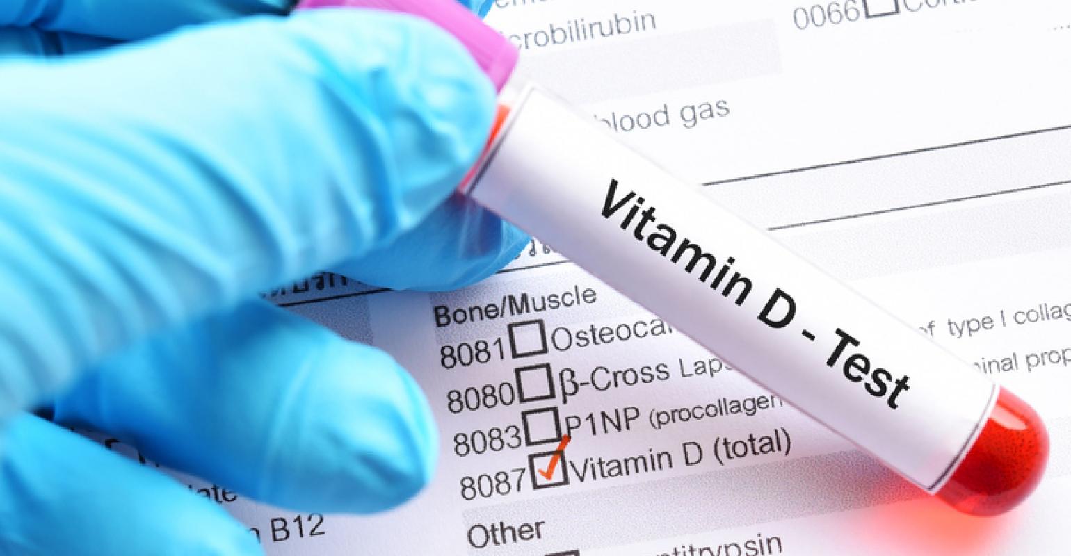
Serum concentration of 25(OH)D is currently the main indicator of vitamin D status. It reflects vitamin D produced endogenously and that obtained from foods and supplements [1]. In serum, 25(OH)D has a fairly long circulating half-life of 15 days [1]. Serum concentrations of 25(OH)D are reported in both nanomoles per liter (nmol/L) and nanograms per milliliter (ng/mL). One nmol/L is equal to 0.4 ng/mL, and 1 ng/mL is equal to 2.5 nmol/L.
Assessing vitamin D status by measuring serum 25(OH)D concentrations is complicated by the considerable variability of the available assays (the two most common ones involve antibodies or chromatography) used by laboratories that conduct the analyses [5,6]. As a result, a finding can be falsely low or falsely high, depending on the assay used and the laboratory. The international Vitamin D Standardization Program has developed procedures for standardizing the laboratory measurement of 25(OH)D to improve clinical and public health practice [5,7-10].
In contrast to 25(OH)D, circulating 1,25(OH)2D is generally not a good indicator of vitamin D status because it has a short half-life measured in hours, and serum levels are tightly regulated by parathyroid hormone, calcium, and phosphate [1]. Levels of 1,25(OH)2D do not typically decrease until vitamin D deficiency is severe [2].
Serum concentrations of 25(OH)D and health
Although 25(OH)D functions as a biomarker of exposure, the extent to which 25(OH)D levels also serve as a biomarker of effect on the body (i.e., relating to health status or outcomes) is not clear [1,3].
Researchers have not definitively identified serum concentrations of 25(OH)D associated with deficiency (e.g., rickets), adequacy for bone health, and overall health. After reviewing data on vitamin D needs, an expert committee of the Food and Nutrition Board (FNB) at the National Academies of Sciences, Engineering, and Medicine (NASEM) concluded that people are at risk of vitamin D deficiency at serum 25(OH)D concentrations less than 30 nmol/L (12 ng/mL; see Table 1 for definitions of “deficiency” and “inadequacy”) [1]. Some people are potentially at risk of inadequacy at 30 to 50 nmol/L (12–20 ng/mL). Levels of 50 nmol/L (20 ng/mL) or more are sufficient for most people. In contrast, the Endocrine Society stated that, for clinical practice, a serum 25(OH)D concentration of more than 75 nmol/L (30 ng/mL) is necessary to maximize the effect of vitamin D on calcium, bone, and muscle metabolism [11,12]. The FNB committee also noted that serum concentrations greater than 125 nmol/L (50 ng/mL) can be associated with adverse effects [1] (Table 1).